Medial Collateral Ligament (MCL) Injuries

The knee looks simple from the outside, but it’s one of the most mechanically complex joints in the human body. It bends, straightens, rotates (slightly), absorbs shock, and supports all of your body weight every time you stand, move, or pivot. Because it has to be both stable and flexible, the knee relies on multiple ligaments and structures to keep everything aligned.

Among those stabilizing structures, the medial collateral ligament (MCL) is one of the most commonly injured. Located on the inner side of the knee, it acts like a firm strap preventing the knee from buckling inward when force is applied from the outside. In fact, MCL injuries account for the largest percentage of all knee ligament injuries and are especially common in sports like skiing, soccer, football, and basketball.1

But MCL injuries aren’t limited to athletes – from a slip on wet flooring to an abrupt twist while stepping off a curb, anyone can sustain one.

Mechanics of an MCL Injury

The MCL stretches from the medial femoral epicondyle (the inner side of the thighbone) to the medial tibial condyle (the top of the shinbone). Its job is to resist valgus stress – the inward force that pushes your knee toward midline.

Most MCL injuries occur when the ligament is overstretched or overloaded:

  • Sports accidents: abrupt cutting, twisting, pivoting, or catching an edge while skiing. Up to 60% of skiing-related knee injuries involve the MCL.1
  • Direct impact: a blow to the outer knee (e.g., a football tackle or a playful dog) that forces the knee inward.
  • Non-sports causes: slipping on ice, sudden knee buckling, or twisting while carrying heavy weight.

Sometimes an MCL tear is the only injury. Other times, it occurs with damage to nearby structures, like in the “unhappy triad” (or terrible triad)- a combination of injury to the MCL, ACL (anterior cruciate ligament), and the medial meniscus.

How to Know the MCL is Injured

MCL injuries can range from mild stretching (Grade I) to complete tears (Grade III), and also may depend on the location of the tear, i.e. midsubstance or pulled off the bone. Symptoms depend on the severity, but the location of the pain is usually a big clue. Common symptoms include:

  • Pain on the inner (medial) side of the knee
  • Swelling or bruising along the inner knee
  • Feeling like the knee might “give way”
  • A popping sensation at the moment of injury
  • Tenderness along the ligament (either near the thighbone, near the shinbone, or along the mid-portion)

If you suspect an MCL injury, follow basic first aid until you can be evaluated:

  • The RICE Method
    • Rest: avoid painful activities
    • Ice: 15 to 20 minutes at a time
    • Compression: wrap lightly to reduce swelling
    • Elevation: above heart level if possible
  • NSAIDs such as ibuprofen can help with swelling and pain (if medically appropriate for you).

If you can’t walk without limping, swelling is significant, your knee feels unstable, pain persists for more than a few days, you heard a “pop” or felt the knee shift, and/or you suspect more than one structure is injured, you need to see a physician.

Inner-knee tenderness can sometimes mimic other issues (like a medial meniscus tear or pes anserine bursitis), so a proper exam is important.

Diagnosis and Treatment of an MCL Injury

A trained clinician can diagnose many MCL injuries just by examining the knee, and getting in to see your physician as close to the time of injury as possible will make for a more accurate diagnosis. Physical assessments and aspects you’ll likely encounter include:

  • Checking for tenderness directly along the ligament
  • Assessing any swelling localized to the inner knee
  • Range of motion measurements
  • Watching for gait changes like limping or guarding the area
  • Evaluating stability under a valgus stress test (pushing the knee inward)

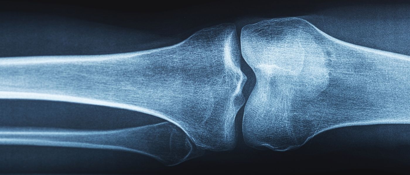
Imaging is used to rule out additional injuries or confirm severity:

  • X-ray: rules out bone injuries, including small fractures. Sometimes it reveals an old MCL injury if a Pellegrini-Stieda lesion (MCL calcification) is present.
  • MRI: the gold standard for viewing ligament tears and detecting associated injuries like ACL or meniscus damage.
  • Ultrasound: a quick, low-cost option that can identify the injury in real time and identify inflammation.

One of the most reassuring things about the MCL is that it has a strong blood supply, which means it heals better than many other knee ligaments. Treatment of the injury depends on the grade.

Grade I is a mild strain that entails a slight stretch, microscopic tear, or minimal instability. Treatment is conservative and typically includes the RICE method, NSAIDs, short-term bracing, and/or early physical therapy focusing on quadriceps strengthening and gradual return to normal activity (usually 10 to 14 days).

Grade II involves more pain and swelling with mild to moderate looseness on valgus stress testing. Treatment is conservative in most cases, and might also include short-term use of a knee brace or immobilizer and/or progressively advancing physical therapy. The recovery timeline varies but is usually several weeks, and returning to normal activities or athletics requires demonstrating equal strength in both legs.

Grade III is a complete tear with significant laxity (looseness) and often occurs with other associated injuries to the ACL or meniscus. Treatment might still verge on conservative if it’s isolated, but surgery to repair or reconstruct using a graft might be recommended for athletes or when there is rotational instability or associated ligament injury. Rehabilitation for a grade III is more intrusive, and can include a hinged brace, early range-of-motion exercises, gradual progression to weight-bearing, and/or closed-chain strengthening exercises (keeping the foot planted). Full return to athletics or normal activities usually takes several months.

MCL injuries generally heal well, and the likelihood of re-injury is low as long as you complete physical therapy and follow your rehabilitation plan. The biggest risks to re-injury involve:

  • Returning to play too early
  • Failing to strengthen supporting muscles
  • Missing associated injuries (ACL or meniscus tears)
  • Developing stiffness from prolonged immobilization
  • Poor adherence to rehab, which can lead to calcification at the injury site (Pellegrini-Stieda lesion)

Fortunately, with accurate diagnosis, proper early care, and a guided rehabilitation program, most MCL injuries heal fully without surgery. Whether you’re an athlete, a weekend warrior, or someone who simply took a bad step, the team at Premier Orthopaedic & Trauma Specialists has your back – and your knees!

  1. Naqvi, U., & Sherman, A. l. (2023, July 17). Medial Collateral Ligament (MCL) Knee Injury. PubMed; StatPearls Publishing. https://www.ncbi.nlm.nih.gov/books/NBK431095/.